Roof Of Fourth Ventricle Anatomy

Pin By Sheena Hunter On College Brain Anatomy Medical Anatomy Medical Knowledge

Pin By Sheena Hunter On College Brain Anatomy Medical Anatomy Medical Knowledge

Brain Atlas Of Human Anatomy With Mri Mri Brain Brain Structure Human Anatomy

Brain Atlas Of Human Anatomy With Mri Mri Brain Brain Structure Human Anatomy

14 3 Brainstem The Medulla Oblongata Relays Signals Between 1278x1500 Png Brain Anatomy Cranial Nerves Nervous System Anatomy

14 3 Brainstem The Medulla Oblongata Relays Signals Between 1278x1500 Png Brain Anatomy Cranial Nerves Nervous System Anatomy

Midbrain5 Jpg 307 397 Basic Anatomy And Physiology Brain Anatomy Medical Anatomy

Midbrain5 Jpg 307 397 Basic Anatomy And Physiology Brain Anatomy Medical Anatomy

14 3 Brainstem The Medulla Oblongata Relays Signals Between The Rest Of The Brain And The Spinal Co Nervous System Anatomy Brain Anatomy Craniosacral Therapy

14 3 Brainstem The Medulla Oblongata Relays Signals Between The Rest Of The Brain And The Spinal Co Nervous System Anatomy Brain Anatomy Craniosacral Therapy

Brain And The Cranial Nerves Cranial Nerves Reticular Formation Nervous System

Brain And The Cranial Nerves Cranial Nerves Reticular Formation Nervous System

Brain And The Cranial Nerves Cranial Nerves Reticular Formation Nervous System

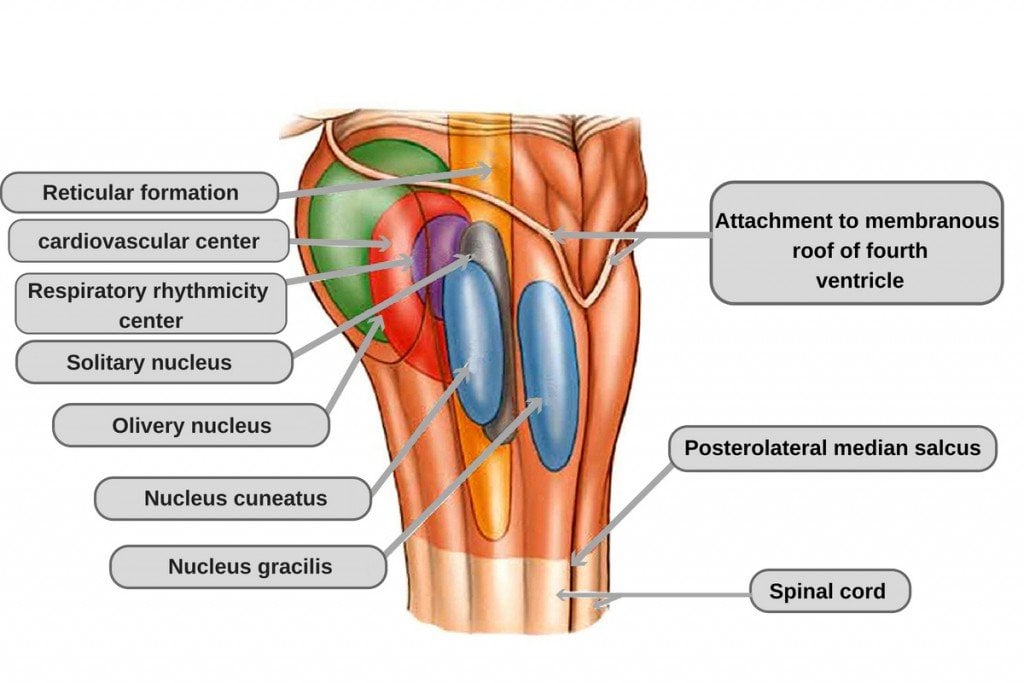
The upper part of the roof is composed by a thin sheet of white matter the superior medullary velum that stretches between both superior cerebellar peduncles.

Roof of fourth ventricle anatomy.

The 4th ventricle is in the form of a spread and irregular cone whose base or floor is supported on the dorsal side of the brainstem. The superior part of the roof is formed by the superior cerebellar peduncles and the superior medullary velum thin sheet of white matter. The upper portion of the roof is formed by the cerebellum. The roof of fourth ventricle is the dorsal surface of the fourth ventricle.

Superior fovea upper end of sulcus limitans widens into a triangular depression. Roof posterior wall the roof of the 4th ventricle is tent shaped and has upper and lower sloping surfaces. The roof of ventricle is diamond shaped and can be divided into superior and inferior parts. In the upper part it is formed by superior medullary velum white matter between the superior cerebellar peduncles.

The obex is the most caudal tip of the fourth ventricle. Lateral to sulcus limitans. Anatomical terms of neuroanatomy. The apex of the tent goes posteriorly into the white core of the cerebellum.

The fourth ventricle has a roof at its upper posterior surface and a floor at its lower anterior surface and side walls formed by the cerebellar peduncles nerve bundles joining the structure on the posterior side of the ventricle to the structures on the anterior side. It corresponds to the ventral surface of the cerebellum. It corresponds to the ventral surface of the cerebellum. The fourth ventricle contains choroid plexus along its roof along the tela choroidea which may protrude out the lateral foramina of luschka.

Medial eminence is bounded laterally by sulcus limitans. The roof is composed of the cerebellum located at the back of the brain and the floor is formed by the rhomboid fossa a depression in the brainstem. It is widest at the level of the pontomedullary junction. The roof of fourth ventricle is tent shaped and is divided into upper and lower part.

The roof of the fourth ventricle has presents a tent like apex at the intersection of it s superior and inferior parts. Overlies the vestib ular nuclei hence it is termed vestibular area. This apex also known as the fastigium extends into the white core of the cerebellum. Features of the floor of fourth ventricle median sulcus.

This ventricle has a roof and a floor.

Mynotes4usmle Speech Language Therapy Speech And Language Speech Therapy

Mynotes4usmle Speech Language Therapy Speech And Language Speech Therapy

Cerebellar Structure Function And Vestibular Disorder Brain Anatomy Cerebellum Anatomy Human Anatomy And Physiology

Cerebellar Structure Function And Vestibular Disorder Brain Anatomy Cerebellum Anatomy Human Anatomy And Physiology

Brainstem Neupsy Key In 2020 Medical School Studying Plexus Products Gross Anatomy

Brainstem Neupsy Key In 2020 Medical School Studying Plexus Products Gross Anatomy

Cross Sectional Anatomy Of The Spinal Cord A Relationships To The Vertebra Meninges And Spinal Nerve B Ste Spinal Cord Anatomy Spinal Cord Spinal Nerve

Cross Sectional Anatomy Of The Spinal Cord A Relationships To The Vertebra Meninges And Spinal Nerve B Ste Spinal Cord Anatomy Spinal Cord Spinal Nerve

Development Of The Spinal Cord A Early Stage Of Development B Intermediate Stage Of Development And C Late Nervous System Gross Anatomy Biology Notes

Development Of The Spinal Cord A Early Stage Of Development B Intermediate Stage Of Development And C Late Nervous System Gross Anatomy Biology Notes

Exam 2 Chapter 14 Diagrams And Labeling Flashcards Flashcards Exam Chapter

Exam 2 Chapter 14 Diagrams And Labeling Flashcards Flashcards Exam Chapter

Pin On Trauma Registrar Anatomy Pics

Pin On Trauma Registrar Anatomy Pics

Protection Of The Brain Medical Massage Dura Mater Epidural Hematoma

Protection Of The Brain Medical Massage Dura Mater Epidural Hematoma

Pin De Erendira Ruiz En Neurologia Google Imagenes Google Y Neurologia

Pin De Erendira Ruiz En Neurologia Google Imagenes Google Y Neurologia

Image Result For Facial Colliculus Facial Nerve Reticular Formation Facial

Image Result For Facial Colliculus Facial Nerve Reticular Formation Facial

Head Ct Scan Procedure Radtechonduty Ct Scan Brain Anatomy Radiography

Head Ct Scan Procedure Radtechonduty Ct Scan Brain Anatomy Radiography

The Diaphragma Sellae Or Sellar Diaphragm Is The Circular Fold Of Dura Mater That Almost Completely Roofs The Fossa Hypophyseos Dura Mater Sphenoid Bone Gland

The Diaphragma Sellae Or Sellar Diaphragm Is The Circular Fold Of Dura Mater That Almost Completely Roofs The Fossa Hypophyseos Dura Mater Sphenoid Bone Gland

Mid Brain Structures Including Commissura Brain Structure Corpus Callosum Brain

Mid Brain Structures Including Commissura Brain Structure Corpus Callosum Brain

Right Aortic Arch Double Aortic Arch And Aberrant Subclavian Artery Obgyn Key Subclavian Artery Ultrasound Ultrasound Sonography

Right Aortic Arch Double Aortic Arch And Aberrant Subclavian Artery Obgyn Key Subclavian Artery Ultrasound Ultrasound Sonography

14 3 Brainstem The Medulla Oblongata Relays Signals Between 1278x1500 Png Brain Anatomy Cranial Nerves Nervous System Anatomy

14 3 Brainstem The Medulla Oblongata Relays Signals Between 1278x1500 Png Brain Anatomy Cranial Nerves Nervous System Anatomy

Pin By Draw It To Know It Medical On Https Drawittoknowit Com Medical School Studying Medical Knowledge Medical Terminology Study

Pin By Draw It To Know It Medical On Https Drawittoknowit Com Medical School Studying Medical Knowledge Medical Terminology Study

Pin De Erendira Ruiz En Neurologia Google Imagenes Google Y Neurologia

Pin De Erendira Ruiz En Neurologia Google Imagenes Google Y Neurologia

3

3

Anatomy Question For Neet Pg Exam Solve This Questions For Complete Demotest Click Http Bit Ly 2hy Online Test Series Online Tests This Or That Questions

Anatomy Question For Neet Pg Exam Solve This Questions For Complete Demotest Click Http Bit Ly 2hy Online Test Series Online Tests This Or That Questions

3vv Three Vessel View Diagnostic Medical Sonography Echocardiogram Medical Ultrasound

3vv Three Vessel View Diagnostic Medical Sonography Echocardiogram Medical Ultrasound

Pin On I See Sound Ob Gyn

Pin On I See Sound Ob Gyn

Ependymoma Of Posterior Third Ventricle This Is A Case Of A Common Paediatric Tumour Being Found In An Uncommon Locat Radiology Radiology Imaging Mri Brain

Ependymoma Of Posterior Third Ventricle This Is A Case Of A Common Paediatric Tumour Being Found In An Uncommon Locat Radiology Radiology Imaging Mri Brain

Source : pinterest.com